The focus on drug discovery has been shifted from the traditional small molecules to biologics and new modalities as a result of increasing research to address unmet medical needs. This evolution requires drug delivery technologies capable of carrying the drug intact to specific sites in the body and across cellular membranes to reach their targets. Engineered nanoparticles have emerged over the past decades as effective and customizable drug delivery platforms.
Due to the versatility of nanoparticulate systems they are defined broadly as materials engineered with at least one dimension or structure in the range of 1–100 nm or as particles with properties related to their nanoscale dimension, even if this dimension is up to 1 μm. In Ardena we define nanoparticles as having a size up to 200 nm. While the concept of entrapping the drug in nanoparticles has been known for many years, the increasing mechanistic understanding and constant advances in optimizing nanoparticulate drug delivery technologies for specific applications continues to accelerate the progress and use in drug product development .
Specific regulatory pathways to stimulate nanoparticle-based drug delivery systems have been drafted by regulatory authorities providing meaningful guidance for research, development, and manufacturing. With the outbreak of the pandemic, the successful development and emergency use of mRNA lipid nanoparticulate vaccines in record time was a quantum leap forward for nanoparticle drug delivery technology. This encouraged drug developers to intensify their investments in engineered nanoparticle delivery for innovative therapeutics approaches.
Introduction
The focus on drug discovery has been shifted from the traditional small molecules to biologics and new modalities as a result of increasing research to address unmet medical needs. This evolution requires drug delivery technologies capable of carrying the drug intact to specific sites in the body and across cellular membranes to reach their targets [1]. Engineered nanoparticles have emerged over the past decades as effective and customizable drug delivery platforms.
Due to the versatility of nanoparticulate systems they are defined broadly as materials engineered with at least one dimension or structure in the range of 1–100 nm or as particles with properties related to their nanoscale dimension, even if this dimension is up to 1 μm [2]. In Ardena we define nanoparticles as having a size up to 200 nm. While the concept of entrapping the drug in nanoparticles has been known for many years, the increasing mechanistic understanding and constant advances in optimizing nanoparticulate drug delivery technologies for specific applications continues to accelerate the progress and use in drug product development [3].
Specific regulatory pathways to stimulate nanoparticle-based drug delivery systems have been drafted by regulatory authorities providing meaningful guidance for research, development, and manufacturing [4]. With the outbreak of the pandemic, the successful development and emergency use of mRNA lipid nanoparticulate vaccines in record time was a quantum leap forward for nanoparticle drug delivery technology [5]. This encouraged drug developers to intensify their investments in engineered nanoparticle delivery for innovative therapeutics approaches.
Characterization of nanoparticle drug delivery systems for pre-clinical and clinical testing
Nanoparticles is an emerging field in biomedical research and drug development. They are developed to overcome the barriers and limitations of traditional drug delivery systems and they can be designed to target the delivery of a drug compound to certain tissues, cells, or intracellular targets, to have reduced toxicity, to prolong systemic exposure, to increase bioavailability, or to overcome other barriers of traditional formulation approaches. Various nanoparticles exist with a broad range of chemistries (e.g., based on lipids, peptides, polymers, metal oxides), architectures (e.g., micelles, vesicles such as liposomes, solid nanoparticles, core-shell like structures), and surface properties.
Their uniqueness is based on their physicochemical properties, especially particle size distribution (PSD) (i.e., average particle size and size polydispersity), surface charge, and structure. These properties can be optimized to enable the entrapped or conjugated drug to be delivered to a specific target or to improve physicochemical stability in the biologic fluids. Consequently nanoparticles are complex by nature because of their multiple characteristics. In addition some specific requirements result from the potential association of nanoparticles with specific adverse reactions like immunological responses such as complement activation-related pseudoallergy (CARPA) and induction of cytokines related to the particle size, zeta potential, and formulation components, which must be carefully considered throughout development [6, 7].
Establishing the critical quality attributes (CQAs) and associated specifications is not trivial. Neither is setting the proper excipient specifications, process and manufacturing conditions to be reproducible for clinical and commercial GMP manufacturing [8]. Me-too generic versions of iron sugar nanoparticles were developed and considered similar to the originator, nevertheless later clinical studies revealed significant differences in efficacy and clinical performance in comparative trials [9].
In drug development a similar challenge exists as the formulation and process are not completely understood and validated in the early phases of drug development. To mitigate the risk a comprehensive, in-depth characterization of engineered particles is essential from early drug development onwards to ensure consistency and an accurate interpretation of the preclinical and clinical results. Such characterization requires a whole range of different analytical methods and experience in selecting the right analytical method for the specific formulation characteristic. The CQAs and the methods to measure them can be divided in 5 categories (Figure 1). The PSD and shape in particular are considered to be CQAs for any nanoparticle as they effect the pharmacokinetics, distribution, interaction with cells, and cellular uptake [10].
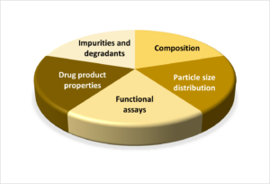
Figure 1. The CQAs of nano-formulations can be categorized in five groups.
From the various analytical methods applied by Ardena for the in-depth characterization of nanoparticles, asymmetric flow field-flow fractionation (AF4) coupled with multi-angle light scattering (MALS) and dynamic light scattering (DLS) has been established for qualitative and quantitative analysis of PSD, molar mass distribution (MMD), shape, and free drug compound analysis for a broad range of different types of nanoparticles.
Asymmetric flow field-flow Fractionation (AF4)
Field-flow fractionation (FFF) is a flow-assisted separation methodology similar to liquid chromatography where the separation takes place inside an open channel under mild conditions [11, 12]. FFF encompasses a family of techniques based on the principle of a thin channel geometry creating a laminar channel flow with a parabolic flow profile and an applied field perpendicular to the channel flow. Over the years FFF systems with different applied force fields (such as thermal, centrifugal, flow, electric or magnetic forces) have been developed to separate biomolecules and nanoparticles according to their specific properties such as size, mass, or charge. In biopharmaceutical sciences the asymmetric flow field-flow fractionation (AF4) has become the major FFF technology used where the applied field is an additional flow, and the separation is solely based on the hydrodynamic size. The AF4 channel consists of a solid top plate with fluid connectors and a bottom plate with a semipermeable membrane placed above a frit (Figure 2). The carrier liquid is allowed to pass through (cross-flow) while the particles are retained, accumulating close to the membrane. Due to their upwards diffusion, the particles are entering different flow streams of the parabolic flow profile and they separate based on their diffusion coefficients and, therefore, based on their hydrodynamic size.
AF4 can fractionate the different subpopulations of the nanoparticles and unbound components based on size for qualitative and quantitative analysis. In contrast to high-performance size-exclusion chromatography (HPSEC), AF4 does not use packing material or chromatographic support exposing the samples to very low pressure and shear forces, which enables labile nanoparticles to be analysed. Other advantages compared to HPSEC are higher sample recovery, operation with nearly all biorelevant buffer media, and a higher upper size limit. AF4 is ISO standardized acknowledged by regulatory authorities as a method for nanoparticle size characterization [13, 14]. It is recognized as a very versatile separation method especially for colloidal systems, such as macromolecules (e.g., proteins, polymers), nanoparticles (based on polymers/lipids/metal oxides/biomolecules) and other complex systems (e.g., blood plasma, viruses). Its application ranges from the early formulation development and characterization, including forced and real time stability testing through to the clinical, and if required to the commercial GMP release testing.
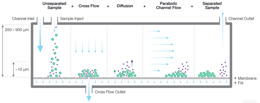
Figure 2. Schematic illustration of the separation mechanism in AF4.
Due to the simultaneous actions of cross flow and diffusion, particles are migrating along the channel with different velocities and separate according to their size; smaller particles move faster because they diffuse into the higher flow velocity region due to their higher diffusion coefficients. [Courtesy from Wyatt https://www.wyatt.com/library/theory/flow-field-flow-fractionation-theory.html]
When coupled online with various detectors AF4 can evaluate multiple CQAs of a complex product sample simultaneously without the need of sample pre-treatment. To determine PSD, MMD, aggregation, or morphology, AF4 coupled with DLS and/or MALS are methods of choice during the lead formulation, process development, and the (pre-)clinical studies. The AF4-MALS/DLS system allows particle size characterization with high resolution even in very complex systems [15]. The PSD obtained by DLS is based on the hydrodynamic size (Rh) and by MALS on the radius of gyration (Rg), which is the root mean square distance of the particles from its center of gravity. This principle favors MALS as a technology to study surface interactions of nanoparticles with endogenous proteins, monitor the change of the particle size and analyse the structure of secondary nanoparticles with adsorbed proteins. In addition indirect information regarding the shape can be achieved from the ratio Rg/Rh [15]: for compact spheres the ratio is 0.77, for empty liposomes 1, and for elongated particles >1.
Application of AF4 in drug development
During the nanoparticle pre-formulation preliminary characterization of the PSD by (batch mode) DLS or nanoparticle tracking analysis (NTA) can serve the purpose of selecting the lead formulation candidate (Figure 3). Although DLS and NTA are suitable to measure samples with a very narrow size distribution, the results are less accurate for samples with broader size distributions and should serve only for batch-to-batch comparison purposes. In addition DLS has low resolution (cannot discriminate between monomer and dimer) and NTA measures only a very small fraction of the sample. Transmission electron microscopy (TEM) is a great tool to visualize the nanoparticles but it requires sample preparation that may alter the size, the images are only two dimensional, and it can estimate PSD based in a very small number of particles. For the preclinical studies an in-depth orthogonal, high resolution nanoparticle analysis, including the evaluation of their fate in relevant biologic fluids is essential to develop a critical understanding of the pharmacology and toxicology of the designed nanoparticles [15].
Figure 3. Particle size characterization during nanoparticle formulation and process development.
Phase appropriate scaling from comparative sizing with DLS/NTA to full scale characterization of size and aggregation/interaction in standard media and biorelevant media after separation from interacting protein.
Multiple analytical methods can be applied to the fractionated subpopulations. Online MALS and DLS detectors are used for the determination of PSD, MMD, morphology, free components, shape, nanoparticle stability (aggregation), and protein binding in biologic fluids [16, 17]. Online UV detection can be used for the determination of a single component concentration along with the particle size measurement [18]. Furthermore many other analytical technologies can be coupled offline with AF4 by collecting fractions for further analysis to investigate if there are size-dependent differences in their zeta potential, drug content, purity, stability, or surface structure [19].
At Ardena we have an AF4 system coupled online with RI, UV, MALS and DLS detectors. The system is also connected to a fraction collector which enables offline coupling with various other techniques. We understand the versatility of AF4 as a separation technology and leverage the strength of each analytical method for the in-depth nanoparticle characterization by selecting the most meaningful scientific product-specific approach for each nanoparticle according and appropriate to the stage of the development.
References
- McGoron AJ (2020) Perspectives on the Future of Nanomedicine to Impact Patients: An Analysis of US Federal Funding and Interventional Clinical Trials. Bioconjugate Chem. 2020, 31(3): 436-447
- Mühlebach S (2018) Regulatory challenges of nanomedicines and their follow-on versions: A generic or similar approach? Adv Drug Del Rev 131: 122–131
Mitchell et al (2021) Engineering precision nanoparticles for drug delivery. Nature Rev Drug Discov 20: 101-124 - Halamoda-Kenzaoui et al (2019) Anticipation of regulatory needs for nanotechnology enabled health products. Publications Office of the European Union, Luxembourg, doi:10.2760/596822, JRC118190
- Schoenmaker et al (2021) mRNA-lipid nanoparticle COVID-19 vaccines: Structure and stability. Int J Pharm 601: 120586
- Clogston JD (2021) The importance of nanoparticle physicochemical characterization for immunology research: What we learned and what we still need to understand. Adv Drug Del Rev 176: 113897
- Sellaturay et al (2021) Polyethylene glycol (PEG) is a cause of anaphylaxis to the Pfizer/BioNTech mRNA COVID-19 vaccine. Clin Exp Allergy 51(6): 861-863.
- Truong et al (2021) Microfluidic generated immunomodulatory nanoparticles and formulation dependent effects on lipopolysaccharide induced macrophage inflammation. The AAPS Journal 24(1): 6
- Rottembourg et al (2011) Do two intravenous iron sucrose preparations have the same efficacy? Nephrol Dial Transplant. 26(10): 3262–3267
- Hoshyar et al (2016) The effect of nanoparticle size on in vivo pharmacokinetics and cellular interaction. Nanomed 11(6): 673–692.
- Contado C (2017) Field-flow fractionation techniques to explore the “nano-world”. Anal Bioanal Chem 409: 2501–2518
- Giddings et al (1993) Field-flow fractionation: Analysis of macromolecular, colloidal, and Particulate Materials. Science 260(5113): 1456-1465
- ISO 21362 (2018) Nanotechnologies — Analysis of nano-objects using asymmetrical flow and centrifugal field-flow fractionation
- Gioria et al (2018) Are existing standard methods suitable for the evaluation of nanomedicines: some case studies. Nanomed 13(5): 539-554
- Caputo et al (2019) Measuring particle size distribution by asymmetric flow field-flow fractionation: A powerful method for the preclinical characterization of lipid-based nanoparticles. Mol. Pharmaceutics 16: 756-767
- Caputo et al (2019) Measuring particle size distribution of nanoparticle enabled medicinal products, the joint view of EUNCL and NCI-NCL. A step-by-step approach combining orthogonal measurements with increasing complexity. J Contr Rel 299: 31-43
- Ventouri et al (2022) Field-flow fractionation for molecular-interaction studies of labile and complex systems: A critical review. Anal Chim Acta 1193: 339396
- Klein et al (2020) Advanced nanomedicine characterization by DLS and AF4-UV-MALS: Application to a HIV nanovaccine. J Pharm Biomed Anal 179: 113017
- Ansar et al (2020) Characterization of doxorubicin liposomal formulations for size-based distribution of drug and excipients using asymmetric-flow field-flow fractionation (AF4) and liquid chromatography-mass spectrometry (LC-MS) Int J Pharm 574: 118906